Theme C: Mechanically stimulable luminescence and photostimulable mechanical properties
Mechanofluorochromism
Within the framework of the ERC project MECHANO-FLUO (2017-2023), we are developing the study of mechanofluorochromic molecules and materials in four directions:
a) Development of new mechanofluorochromic compounds
The objectives are on the one hand to establish relationships between the structure of a compound and its mechanofluorochromic properties, and on the other hand to obtain efficient materials for the other parts of the project. For this purpose, we have developed a small library of compounds, either synthesized at PPSM (boron-diketone complexes, polydiacetylenes), or studied in collaboration (pyrene derivatives - coll. J. Zakrewski, Lodz, Poland; rhenium complexes - coll. S. Fery-Forgues, Toulouse; iiphenylamine derivatives - coll. J. Roncali, Moltech Angers) in order to obtain compounds with different responses to different mechanical stresses (pressure, shear), sensitivities and reversibilities. The study of materials coupling mechanofluorochromism to circularly polarised luminescence (CPL - coll. T. Kawai, NAIST, Japan) is also being developed.
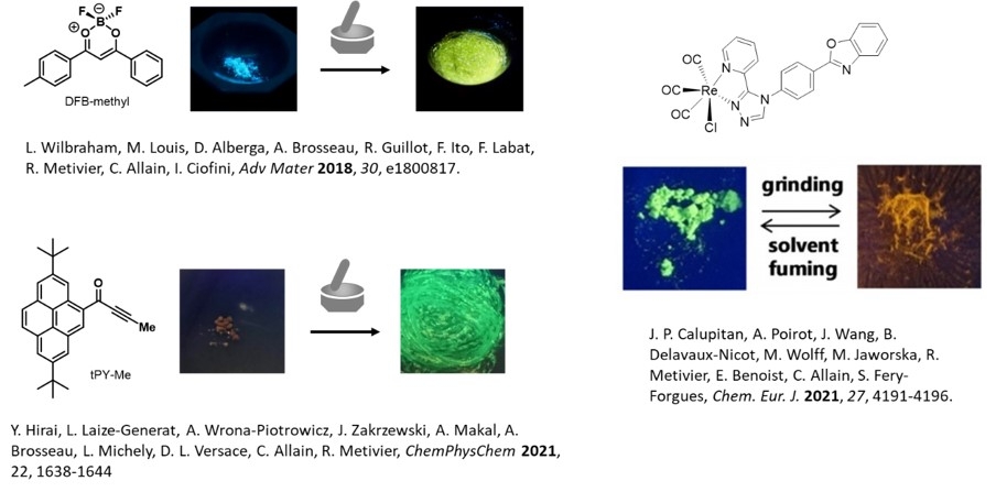
Examples of mechanofluorochromic compounds recently studied in PPSM. The pictures show the luminescence before and after grinding in a mortar
b) Study of mechanofluorochromism at the nanometric scale
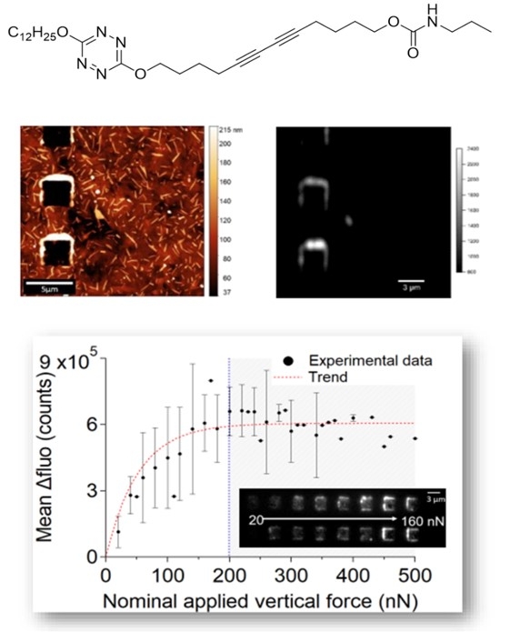
Fluorescence microscopy coupled to AFM is our preferred instrumentation here. We have demonstrated a mechanofluorochromic response at the nanometric scale on three families of compounds. In one case, we demonstrated a different response from that observed at the macroscopic scale (J. Phys. Chem. Lett 2019) and in the other two cases, which were on-off systems, we were able to show an increase in fluorescence as the force applied to the material with the AFM tip increased (Chem. Commun. 2019, Adv. Mater. Interfaces 2022). This study will be continued on other compounds, but especially developed to better quantify the response at the nanometric scale and to move towards a measurement of the fluorescence emission during the application of a force as well as towards time-resolved fluorescence measurements under the microscope (see theme instrumentation and innovative set ups).
Example of the tetrazine-diacetylene mechanofluorochrome TzDA1 dyad (top), TzDA1 thin film obtained by vacuum evaporation after application of a mechanical force by AFM in contact mode observed by topographic AFM (middle left) and by fluorescence microscopy (middle right) and increase of the fluorescence signal as a function of the vertical force applied by the AFM tip (bottom)
c) Quantification of mechanofluorochromism on a macroscopic scale (coll. L. Bodelot, LMS, École Polytechnique)
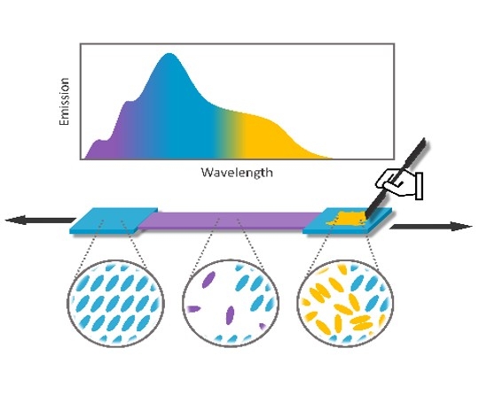
On the one hand, we sought to quantify (nature and intensity) the force to be applied to a molecular material that lead to a change in fluorescence, in order to guide our molecular engineering work (cf. a). For this purpose, L. Bodelot designed a set-up allowing controlled application of compression or shear to a material in powder form. A detailed study on the compound DFB-Me showed a significantly higher sensitivity to shear stresses than to compressive stresses (J. Mater. Chem. C 2021), which seems to be confirmed by preliminary results on other structurally different compounds. On the other hand, we have developed mechanofluorochromic polymers by non-covalent incorporation of diketone ligand boron complexes into an LLDPE matrix. These polymers showed a different response to traction (appearance of a violet fluorescence) and friction (appearance of a yellow fluorescence), which constitutes a unique behaviour to our knowledge (Macromol. Rapid Commun, 2022). These studies will be continued, in particular by looking at the covalent incorporation of mechanophores into polymers, in order to determine whether such materials can be useful tools for mechanical stress metrology.
Schematic representation of the emissive species involved in the fluorescence of an LLDPE/DFB composite polymer. Before mechanical stress, the fluorophore is crystallized in the polymer matrix (cyan blue fluorescence); after traction the fluorescence of the fluorophore dispersed in the matrix increases (violet emission); after friction the fluorophore crystals are destroyed to form amorphous aggregates (yellow fluorescence)
d) Application in mechanobiology
Our objective is to evaluate whether the compounds developed in a) and characterised at the nanometric scale in b) can be exploited as stress probes in mechanobiology at the cell scale. Thus, we are working on the development of mechanofluorochromic surfaces and we propose (collab. S. Bensalem, B. Le Pioufle, LUMIN, ENS Paris Saclay) to develop microfluidic channels functionalized by the same strategy in which (collab. F. Lopes, LGPM, Centrale Supélec) we will study the passage of microalgae. Indeed, the teams of F. Lopes and B. Le Pioufle's teams have shown that the passage of microalgae in microfluidic channels comprising restrictions facilitates the extraction of compounds of interest produced by these "microfactories", it would therefore be particularly interesting to be able to quantify these forces. More generally, a material that is sensitive enough to respond to the mechanical forces exerted by a cell would be a valuable tool for mechanobiology studies.
Photomechanical systems (R. Métivier, C. Allain, G. Laurent, A. Brosseau)
The second part of this theme concerns the study of photomechanical effects at the nanometric scale. The mechanical response of organic nanosystems subjected to light irradiation is a very attractive and relatively unexplored field of research. Photochromic organic micro- and nanomaterials will be prepared from diarylethenes with long aliphatic side chains to flexibilise the solid (coll. Prof. C. Bertarelli, Politecnico di Milano) or supramolecular solids formed from diarylethenes and elastomeric chains (coll. Dr S. Aloïse, LASIR, Univ. Lille). Illuminated under a microscope and observed by AFM, the effects of expansion, contraction or deformation of the objects will be quantified. Ultimately, the aim is to produce objects capable of imparting a detectable and quantifiable local stress to their environment in the form of a force applied to an AFM tip for example (see theme instrumentation and innovative set ups). The magnitude of this force and the dissipation speed of the mechanical energy triggered by the light will be two key parameters of the system. They will allow us to evaluate the potential application of these systems as nanomachines in the field of nanotechnology, or as nanosources of pressure to provoke a spatially localised shock wave (in a cell for example).
National collaborations
- Dr. Régis Guillot (ICMMO, Université Paris-Saclay)
- Dr. Ilaria Ciofini, Dr. Frédéric Labat (ChimieParisTech)
- Dr. Laurence Bodelot (Ecole Polytechnique)
- Dr. Clément Cabanetos, Dr. Jean Roncali (Moltech Anjou)
- Dr. Suzanne Fery-Forgues (Université Paul Sabatier, Toulouse)
- Dr. Sakina Bensalem, Pr. Bruno Le Pioufle (LUMIN, Institut d’Alembert, ENS Paris-Saclay), Dr. Filipa Lopes (LGPM, Centrale Supélec).
- Dr S. Aloïse, (LASIRE, Univ. Lille)
International collaborations
Research contracts
ERC Mechano-fluo (2017-2023)
D’Alembert Institute project MecaFluoAlgues (2020-2021)
Theme C managers


Participants

Former members
- Dr. Rajesh Bisht
- Dr Benjamin Poggi (read his PhD online)
- Dr Marine Louis (read her PhD online)
