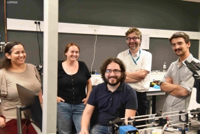
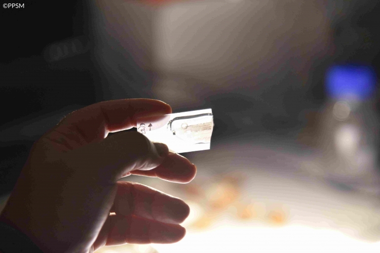
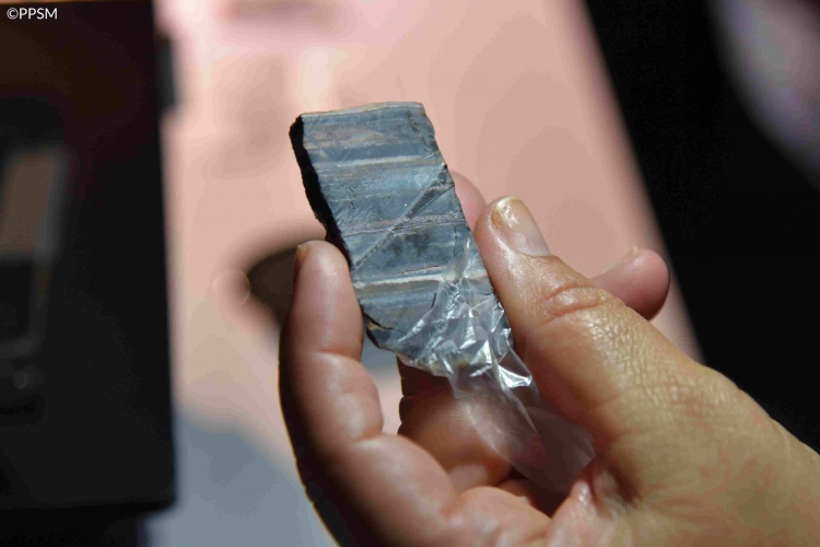
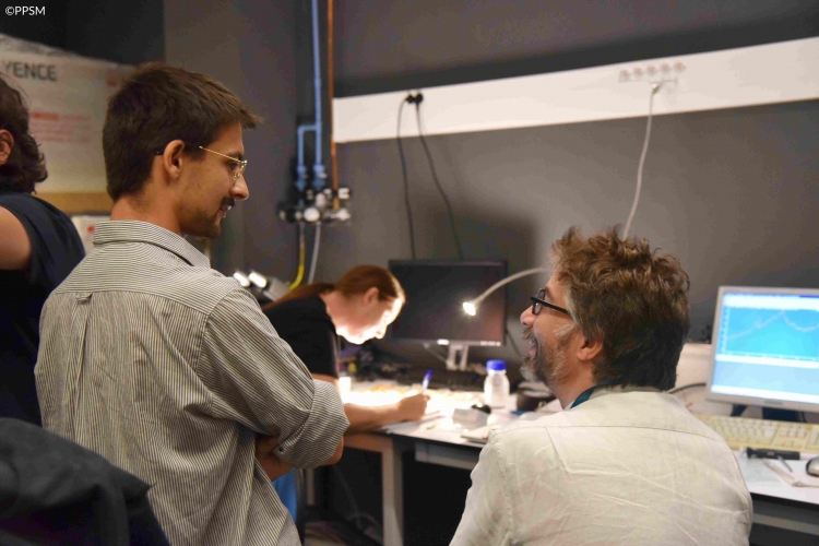
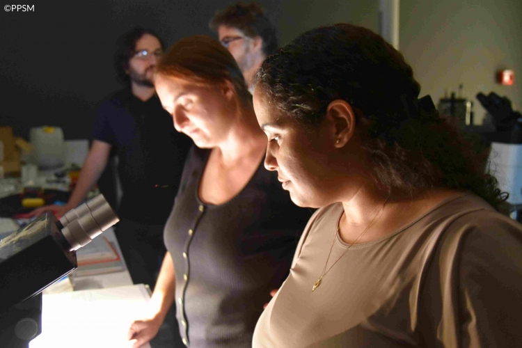
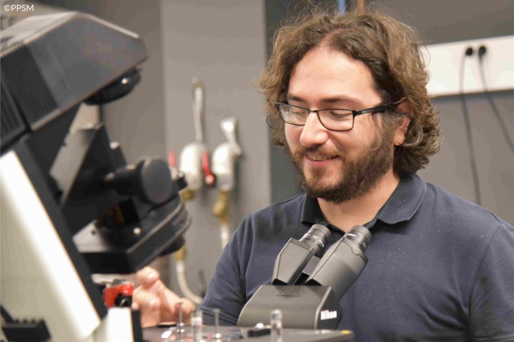
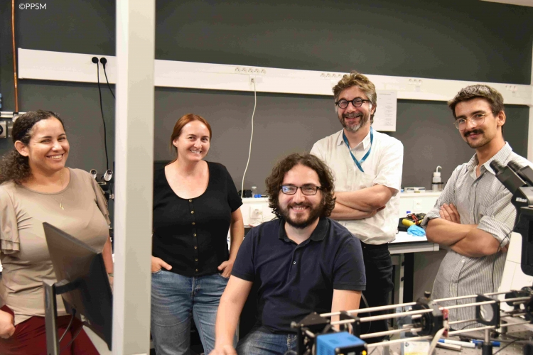
Dr Flavia Callefo and Dr Veronica Teixeira (Brazilian Synchrotron Light Laboratory LNLS, Brazilian Research Center in Energy and Materials CNPEM) are looking for potential microbial biosignatures in extremely old rocks (over 3 billion years old) taken from the Dresser Formation in Australia. During a stay at ENS Paris-Saclay, they used photoluminescence microscopy and Raman microscopy to assess the possible biogenicity of microscopic samples.
Why are scientific stays so important?
The PPSM laboratory regularly welcomes foreign scientists. Such scientific stays are invaluable for fostering partnerships and advancing scientific knowledge because they allow:
- Enhanced exchange of ideas
- Sharing of expertise around specialized equipment and skill development
- Cultural and collaborative exposure
Here, the two Brazilian researchers visited the PPSM laboratory to pursue the collaboration with Dr Loïc Bertrand, in the context of the Collaborative Center Atoms for Heritage recently established by Université Paris-Saclay and the International Atomic Energy Agency, and the starting CNRS 80prime PhD project of Jérémie Ritoux.
A fruitful collaboration between PPSM laboratory and Brazil in the realm of Raman photoluminescence
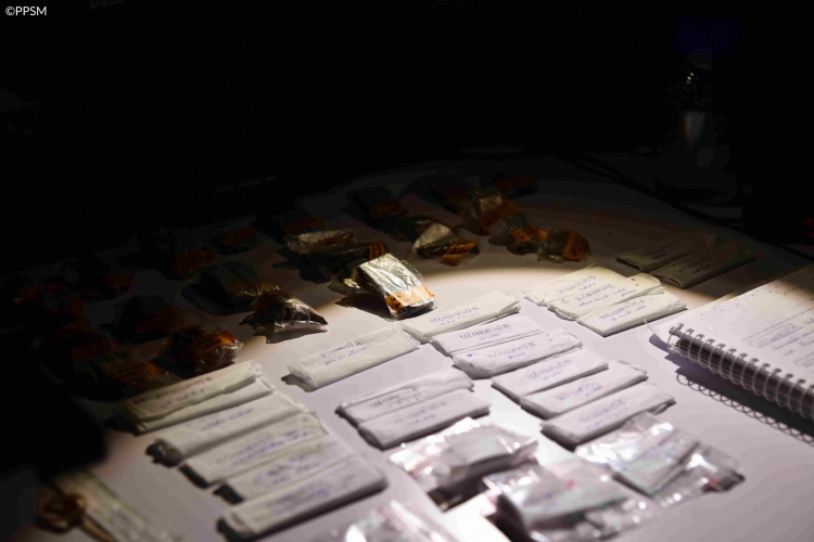
Dr Flavia Callefo and Dr Veronica Teixeira came from Brazil with 25 samples of rocks collected in Dresser Formation, Australia, to exchange and perform experiments with Dr Loïc Bertrand, Dr Vitor Brasiliense, Dr Rémi Métivier and Jérémie Ritoux. The main interest was to analyse some previous selected textures, structures containing putative biominerals in 3 sample sets, each set corresponding to a different location in Dresser Formation.
Using an internally developed instrument, Raman photoluminescence enables researchers to explore the molecular structure and properties of materials by using inelastic light scattering. In other words, it reveals essential information about the chemical composition and structure of materials by analyzing the scattered light when it interacts with samples.
Hence, the collected Raman spectra show characteristic peaks that allowed Dr Flavia Callefo, Dr Veronica Teixeira and their PPSM colleagues to identify different components of the samples and to assess whether they may contain microbial biosignatures.
